Details
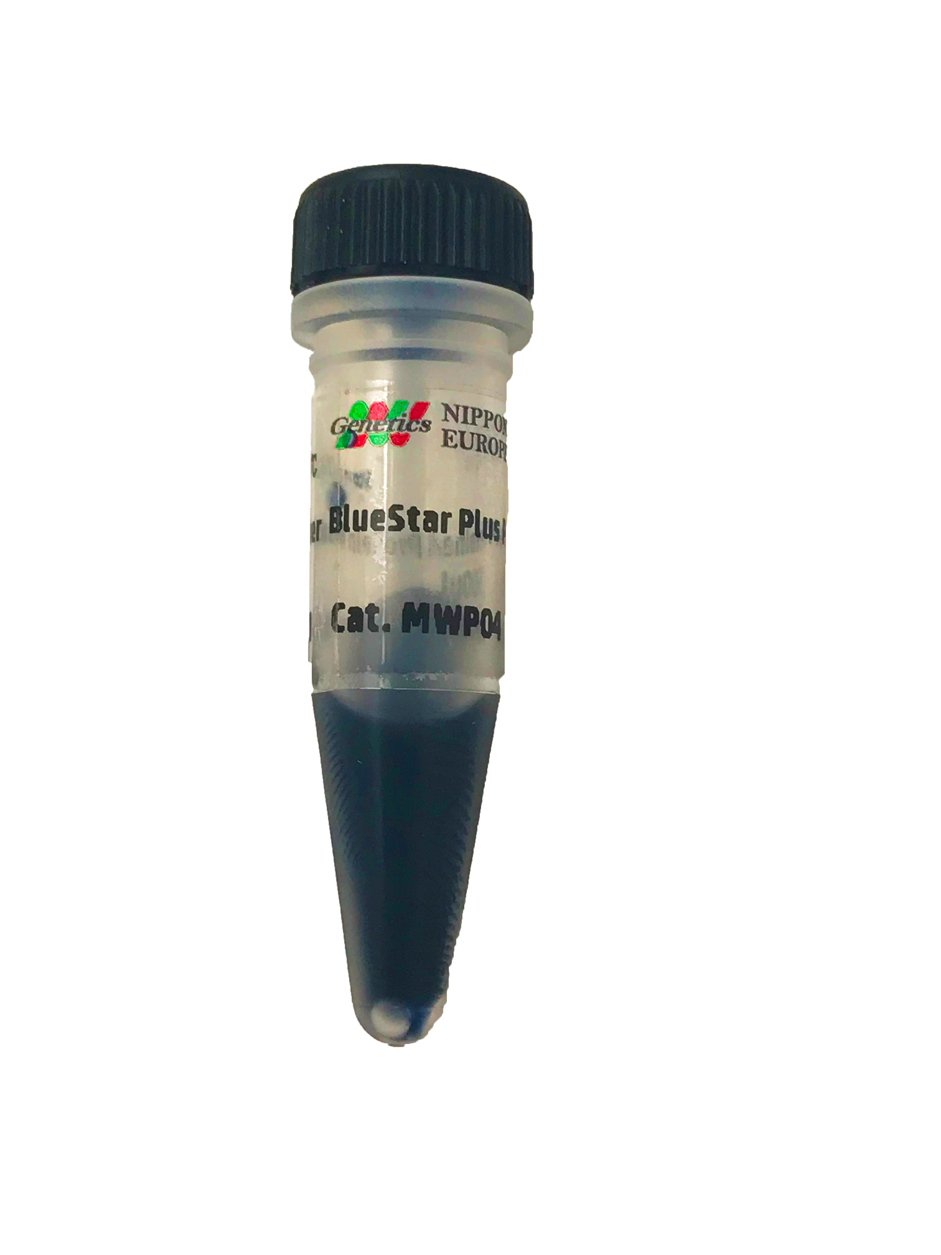
BlueStar PLUS Prestained Protein Marker
The BlueStar PLUS Prestained Protein Marker is a three color protein standard with 12 prestained proteins covering a wide range molecular weights from 10 – 240 kDa.
The Prestained Protein Ladder is designed for monitoring protein separation during SDS-polyacrylamide gel electrophoresis, verification of Western transfer efficiency on membranes (PVDF, nylon, or nitrocellulose) and for approximate sizing of proteins.
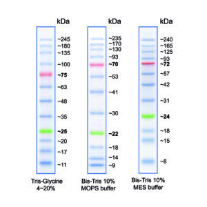
Figure 1: Molecular weight of the marker bands when using different buffer systems.
The molecular weight of the marker bands can vary depending on the gel/buffer system used. This is due to factors of the gel/buffer system that affect the running behaviour of proteins, e.g. migration rate, gel composition, pH-value (see FAQ for more information).
Contents
Approximately 0.1~0.4 mg/ml of each protein in the buffer (20 mM Tris-phosphate, 2 % SDS, 0,2 mM Dithiothreitol, 3.6 M Urea, and 15 % (v/v) Glycerol).
500 µL Protein Marker sufficient for 100 mini gels or 50 large gels.
Storage
Store at -20°C for 24 months.
Stable at +4°C for 3 months.
Stable at +25°C for 2 weeks.
Recommendations for Loading
- Thaw the ladder either at room temperature or at 37-40°C for a few minutes to dissolve precipitated solids. Do not boil.
- Mix thoroughly, to ensure that the solution is homogeneous.
- Load the following volumes of the ladder on SDS-polyacrylamide gel:
– 5 μl per well for mini-gels, 3 μl per well for blots;
– 10 μl per well for large gels, 6 μl per well for blots
Applications
• Monitoring of protein migration during SDS-polyacrylamide gel electrophoresis
• Monitoring of protein transfer onto membranes during Western blotting
• Sizing of proteins on SDS-polyacrylamide gels and Western blots
FAQ
Which amino acids are linked to the BlueStar Plus Prestained Marker MWP04?
Amino acids involved in the dye reaction are those with amine side chain, such as Arg and Lys.
Can the tryptophane been tagged with the trihalo compounds of the biorad gels or the fluorescent dye?
We do not have information regarding the prestained dye interfering with Bio-rad gel systems or the fluorescent dye associated with it.
Does this marker contain Formaldehyde or Formamide?
No.
Why are the molecular weights of the marker bands for the protein markers given separately for different systems?
There are several factors of gel and buffer chemistry that have an influence on the running behaviour of proteins. The most important are:
Migration rate: The migration of proteins in electrophoresis is influenced by the properties of the buffer system, e.g. by the ionic strength, the pH value and the presence of certain ions. The different buffer systems influence the electrophoretic mobility of proteins differently. For example, proteins may migrate faster or slower in one buffer system than in another, resulting in obvious differences in their molecular weight.
Gel composition: The type of polyacrylamide gel and its pore size can influence protein migration. Different buffer systems may be optimised for different gel types, which affects how proteins move through the gel matrix. These differences can lead to variations in the observed molecular weights.
pH: The pH of the electrophoresis buffer can also affect the charge and conformation of the proteins, which in turn affects their migration in the gel. Different buffer systems may have different pH values, leading to variations in molecular weight estimates.
Tris-glycine gels contain a moving front of Cl- ions (from the Tris-HCl in the gel) and negatively charged glycine ions from the running buffer. The glycine ions move slower than the chloride ions through the low pH (pH 6.8) collection gel and form a narrow zone where the proteins are concentrated. When the moving front meets the separating gel with the higher pH (pH 8.8), the glycine ions move quickly past the chloride ions, pulling the proteins through the gel with them, so to speak. Because of this composition, tris-glycine gels work in the strongly alkaline range and reach a pH of up to 9.5 when running.
Only logged in customers may leave a review.


Reviews
There are no reviews yet.